Abdomen Anatomy Female Bowel - Intestines Anatomy Picture Function Location Conditions : The intestines are located inferior to the stomach in the abdominal body cavity.
byAdmin-
0
Abdomen Anatomy Female Bowel - Intestines Anatomy Picture Function Location Conditions : The intestines are located inferior to the stomach in the abdominal body cavity.. The small intestine is a convoluted tube connecting the stomach with the large intestine. Anatomy of pregnancy 12 photos of the anatomy of pregnancy anatomy and pathophysiology of ectopic pregnancy, anatomy and physiology of ruptured ectopic pregnancy, anatomy of pregnancy induced hypertension, anatomy of pregnancy pdf, anatomy when pregnancy, human anatomy, anatomy and pathophysiology of ectopic pregnancy. Abdominal computed tomography (ct) is a type of medical imaging procedure used to diagnose and monitor internal stomach issues, like cancer, bowel obstruction, and abdominal pain. Radiographers suggest an abdominal ct scan to look for the following: Stomach anatomy and its parts.
Abdominal adhesions are the most common cause of obstruction of the small intestine. Abdominal computed tomography (ct) is a type of medical imaging procedure used to diagnose and monitor internal stomach issues, like cancer, bowel obstruction, and abdominal pain. The stomach into the duodenum, which is the tube that leads from the stomach into the intestines. In women, the lowest portion of the abdomen is actually the pelvis and involves the uterus,. The photo of stomach and large intestine is on the woman's body against gray background, people with stomach ache problem concept, female anatomy intestines black icon, vector sign on isolated background.
The Radiology Assistant Us Of The Gi Tract Normal Anatomy from radiologyassistant.nl However, the small intestine is present at the central part and lower parts of the abdominal proper cavity where the large intestine surrounds it. At the level of the pelvic bones, the abdomen ends and the pelvis begins. Those organs include the stomach, small intestine, colon, liver, gallbladder, spleen, and pancreas. • the descending colon travels down the left abdomen. This portion of the small intestine received its name due to its size; The major organs of the abdomen include the. Intestinal obstruction is the partial or complete blockage of the movement of food, fluids, air, or stool through the intestines. This medical exhibit diagram illustrates the anatomy of the female abdomen and pelvis from an anterior front cut away view showing elements of the digestive system the liver stomach and abdominal contents are clearly identified and labeled including the cecum ascending colon transverse colon descending colon and small intestine the image also shows the pelvis uterus and urinary bladder the pelvic bones and femur bones are ghosted beneath the skin
It is approximately 5 meters long and includes the duodenum, jejunum, and ileum.
The small intestine is situated between the stomach and. If you plan to enter a healthcare profession such as nursing, this is something you'll use on the job when performing abdominal assessments (and while documenting). Abdomen anatomy female bowel / 12 920 large intestine stock photos pictures royalty free images istock. The right ovary and fallopian tube are also located in the right lower quadrant in females. In women the lowest portion of the abdomen is actually the pelvis and involves the uterus fallopian tubes and ovaries. Stomach anatomy and its parts. This medical exhibit diagram illustrates the anatomy of the female abdomen and pelvis from an anterior front cut away view showing elements of the digestive system the liver stomach and abdominal contents are clearly identified and labeled including the cecum ascending colon transverse colon descending colon and small intestine the image also shows the pelvis uterus and urinary bladder the pelvic bones and femur bones are ghosted beneath the skin 1 the duodenum can be separated into. It is about 6 meters (20 feet) long and extends from the pylorus of the stomach to the ileocecal junction. It is in fact very difficult to identify the cause of an abdominal pain because of the multiple organs within. The abdomen contains all the digestive organs, including the stomach,. However, the small intestine is present at the central part and lower parts of the abdominal proper cavity where the large intestine surrounds it. This medical exhibit diagram illustrates the anatomy of the female abdomen and pelvis from an anterior front cut away view showing elements of the digestive system.
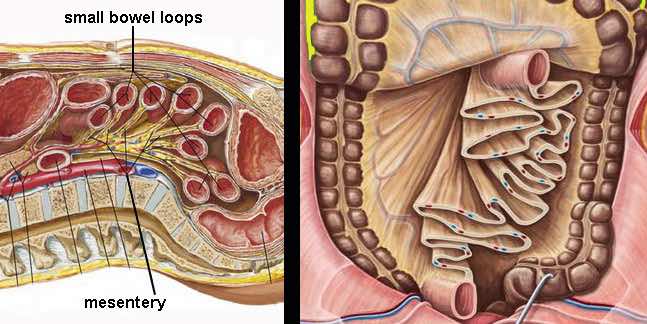
Labeled structures include the large bowel (colon or large intestine), umbilicus, small intestine, ovary, fallopian tube, uterus and bladder. The space below contains the bladder, rectum, and part of the descending colon. It consists of the duodenum , jejunum and ileum.the terminal ileum opens to the cecum of the large intestine at the ileocecal junction ,. The diaphragm forms the upper surface of the abdomen. The right ovary and fallopian tube are also located in the right lower quadrant in females.
Stock Female Pelvis Normal Anatomy Illustrated Verdict from images.squarespace-cdn.com Stomach anatomy and its parts. The small bowel contains prominent mucosal folds known as plicae circulares or valvular connvinetes. The space below contains the bladder, rectum, and part of the descending colon. This portion of the small intestine received its name due to its size; Those organs include the stomach, small intestine, colon, liver, gallbladder, spleen, and pancreas. • the sigmoid colon is a short curving of the colon, just before the rectum. • the descending colon travels down the left abdomen. The major organs of the abdomen include the.
The right ovary and fallopian tube are also located in the right lower quadrant in females.
Various folds or reflections of the peritoneum connect viscera to the abdominal walls or to one another. It is the region where most digestion and. • the descending colon travels down the left abdomen. This medical exhibit diagram illustrates the anatomy of the female abdomen and pelvis from an anterior front cut away view showing elements of the digestive system the liver stomach and abdominal contents are clearly identified and labeled including the cecum ascending colon transverse colon descending colon and small intestine the image also shows the pelvis uterus and urinary bladder the pelvic bones and femur bones are ghosted beneath the skin At the level of the pelvic bones, the abdomen ends and the pelvis begins. The small intestine is divided into. It is about 6 m in length and extends from the pyloric sphincter to the ileocecal junction. Those organs include the stomach, small intestine, colon, liver, gallbladder, spleen, and pancreas. The abdomen contains all the digestive organs, including the stomach,. The space below contains the bladder, rectum, and part of the descending colon. In anatomy and physiology, you'll learn how to divide the abdomen into nine different regions and four different quadrants. This landmark begins at the level of the sacral promontory posteriorly and the pubic symphysis anteriorly. Related posts of anatomy of the abdomen women anatomy of pregnancy.
Those organs include the stomach, small intestine, colon, liver, gallbladder, spleen, and pancreas. In latin, duodenum translates to 12 fingers, which is the approximate length of the organ. Intestinal obstruction is the partial or complete blockage of the movement of food, fluids, air, or stool through the intestines. It is a long tubelike organ that removes water from digested food. The human abdomen is that part in the front of our body between the chest and the waist line.
Anatomy Of The Female Abdomen And Pelvis Cut Away View Doctor Stock from m.psecn.photoshelter.com It is about 6 meters (20 feet) long and extends from the pylorus of the stomach to the ileocecal junction. The colon is further divided into: The intestines are located inferior to the stomach in the abdominal body cavity. If you plan to enter a healthcare profession such as nursing, this is something you'll use on the job when performing abdominal assessments (and while documenting). The jejunum has the most developed and highest concentration. Radiographers suggest an abdominal ct scan to look for the following: National library of medicine was used as the basis to build an exemplary model of the female abdomen analyzing the normal anatomy we found several variations and pathologies of the vhf, such as missing muscles (gemellus superior, psoas. The human abdomen is that part in the front of our body between the chest and the waist line.
Stomach anatomy and its parts.
The major organs of the abdomen include the. Various folds or reflections of the peritoneum connect viscera to the abdominal walls or to one another. Anatomy of pregnancy 12 photos of the anatomy of pregnancy anatomy and pathophysiology of ectopic pregnancy, anatomy and physiology of ruptured ectopic pregnancy, anatomy of pregnancy induced hypertension, anatomy of pregnancy pdf, anatomy when pregnancy, human anatomy, anatomy and pathophysiology of ectopic pregnancy. In women the lowest portion of the abdomen is actually the pelvis and involves the uterus fallopian tubes and ovaries. • the descending colon travels down the left abdomen. The right ovary and fallopian tube are also located in the right lower quadrant in females. It is approximately 5 meters long and includes the duodenum, jejunum, and ileum. The liver stomach and abdominal contents are clearly identified and labeled including the cecum ascending colon transverse colon descending colon and small. This medical exhibit diagram illustrates the anatomy of the female abdomen and pelvis from an anterior front cut away view showing elements of the digestive system. At the level of the pelvic bones, the abdomen ends and the pelvis begins. The colon is further divided into: The liver, stomach, and abdominal contents are clearly identified and labeled, including the cecum, ascending colon, transverse colon, descending colon, and small intestine. The stomach is responsible for the secretion of digestive enzymes and gastric acid required to digest food products.
However, the small intestine is present at the central part and lower parts of the abdominal proper cavity where the large intestine surrounds it abdomen anatomy-female. The abdomen contains all the digestive organs, including the stomach,.